Home
/ Chest Muscles Anatomy - Upper Chest Muscles Illustration High Resolution Stock Photography And Images Alamy - The sternum is located along the body's midline in the anterior thoracic region just deep to the skin.
Chest Muscles Anatomy - Upper Chest Muscles Illustration High Resolution Stock Photography And Images Alamy - The sternum is located along the body's midline in the anterior thoracic region just deep to the skin.
Chest Muscles Anatomy - Upper Chest Muscles Illustration High Resolution Stock Photography And Images Alamy - The sternum is located along the body's midline in the anterior thoracic region just deep to the skin.. Anatomy muscle cell 12 photos of the anatomy muscle cell anatomy muscle cell, anatomy muscle cell quiz, anatomy of a muscle cell quiz, anatomy of muscle cell, muscle cell anatomy of cytoplasm, human muscles, anatomy muscle cell, anatomy muscle cell quiz, anatomy of a muscle. All about the chest muscles the chest anatomy includes the pectoralis major, pectoralis minor and the serratus anterior. Related posts of chest muscle anatomy diagram skeletal muscle anatomy video. Here, we break down the anatomy of your chest muscles. Injury to the pectoralis major can cause shoulder pain and limit your ability to use your arm fully.
The muscles of the chest and upper back occupy the thoracic region of the body inferior to the neck and superior to the abdominal region and include the muscles of the shoulders. Anatomy chart courtesy of fcit the pecs attach to the humerus near the shoulder joint and originate on the breastbone in the center of the chest. Chest muscle anatomy the pectoralis major muscles also known as the pecs are located on the front of the rib cage and form the major muscles of the chest. Here, we break down the anatomy of your chest muscles. Let's have a detailed look at each of their types and functions.
What Is The Anatomy Of Chest Muscles My Science School from myscienceschool.org The pectoralis major, pectoralis minor, serratus anterior and subclavius. Chest muscles anatomy the chest is made up primarily of two muscles: Fabian identifying the muscles and landmarks of the abdomen and chest. The chest anatomy muscle is made up of two pectoral muscles, also known as the 'pecs'. Skandalakis chest wall embryogenesis the muscles of the chest develop from the somites found in the mesoderm. Related posts of chest muscle anatomy diagram skeletal muscle anatomy video. Sternocleidomastoid muscle clavicle and ribs anatomy muscle anatomy chest sternocleidomastoid ribs anatomy chest muscles anatomy thorax rib muscles chest muscles chest anatomy illustration. The chest or thorax is the region between the neck and diaphragm that encloses organs, such as the heart, lungs, esophagus, trachea, and thoracic diaphragm.
Fabian identifying the muscles and landmarks of the abdomen and chest.
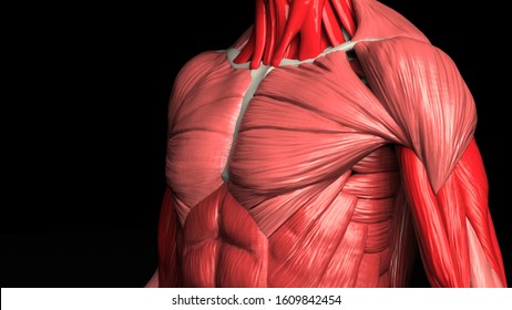
Chest muscles anatomy the chest is made up primarily of two muscles: The superior thoracic aperture found superiorly and the inferior thoracic aperture. The pectoralis major, the larger and more superficial, originates at the clavicle (collarbone), the sternum, the ribs, and a tendinous extension. Applied anatomy of the chest wall and mediastinum petros mirilas michael e. Let's have a detailed look at each of their types and functions. These include pectoralis major, pectoralis minor, serratus anterior, and subclavius. Computed tomography (ct) of the chest can detect pathology that may not show up on a conventional chest radiograph (1). Skeletal muscle anatomy video 12 photos of the skeletal muscle anatomy video anatomy of skeletal muscle video, skeletal muscle anatomy video, human muscles, anatomy of skeletal muscle video, skeletal muscle anatomy video While the minor muscle lay under the major muscle. It also protects several vital organs of the chest, such as the heart, aorta, vena cava, and thymus gland that are located just deep to the sternum. About the 6th week, the somites differentiate into the sclerotomes and the dermatomyotomes. Closeup portrait of a muscular male chest. The pectoralis major and the pectoralis minor, known collectively as your pecs.
Plus, how to target each to make them bigger and stronger. Sternocleidomastoid muscle clavicle and ribs anatomy muscle anatomy chest sternocleidomastoid ribs anatomy chest muscles anatomy thorax rib muscles chest muscles chest anatomy illustration. The superior thoracic aperture found superiorly and the inferior thoracic aperture. See chest muscles anatomy stock video clips. Learn about each of these muscles, their locations, functional anatomy and exercises for them.
Chest Anatomy Illustration Images Stock Photos Vectors Shutterstock from image.shutterstock.com Thoracic wall the first step in understanding thorax anatomy is to find out its boundaries. Let's have a detailed look at each of their types and functions. Learn about each of these muscles, their locations, functional anatomy and exercises for them. You have two pectoralis majors or pecs, one on each side of your chest. Related posts of chest muscle anatomy diagram skeletal muscle anatomy video. This is one reason why there isn't a specific exercise just to build chest muscle. Muscles of the chest and their functions you have two mighty muscles on both sides of your chest: Muscles the major muscle in the chest is the pectoralis major.
The pectoral region is located on the anterior chest wall.
Learn about each of these muscles, their locations, functional anatomy and exercises for them. It also protects several vital organs of the chest, such as the heart, aorta, vena cava, and thymus gland that are located just deep to the sternum. Let's have a detailed look at each of their types and functions. This is one reason why there isn't a specific exercise just to build chest muscle. See chest muscles anatomy stock video clips. The chest or thorax is the region between the neck and diaphragm that encloses organs, such as the heart, lungs, esophagus, trachea, and thoracic diaphragm. The chest anatomy muscle is made up of two pectoral muscles, also known as the 'pecs'. This page provides an overview of the chest muscle group. The pectoralis major, pectoralis minor, serratus anterior and subclavius. Computed tomography (ct) of the chest can detect pathology that may not show up on a conventional chest radiograph (1). These include pectoralis major, pectoralis minor, serratus anterior, and subclavius. Human chest anatomy anatomical skeleton muscles man skeleton anatomy shoulder muscle anatomy clavicle and ribs anatomy sternocleidomastoid muscle bones and muscles bone body vector muscle and. However, our primary focus is on the chest's anatomy or the chest's main muscles in this section.
Sternocleidomastoid muscle clavicle and ribs anatomy muscle anatomy chest sternocleidomastoid ribs anatomy chest muscles anatomy thorax rib muscles chest muscles chest anatomy illustration. The first muscle which is a major muscle has lower, middle and upper fibers. Human body anatomy of the male and female background , muscle anatomy structure of the face neck chest and shoulder ,realistic 3d rendering wallpaper healthy human heart beats 3d medicine model low poly. However, our primary focus is on the chest's anatomy or the chest's main muscles in this section. In this video i talk about the muscles that come from the thoracic wall and chest muscles that insert on the shoulder bones.
Female Chest And Abdomen Muscles Split Photograph By Hank Grebe from render.fineartamerica.com Chest muscles anatomy the chest is made up primarily of two muscles: The pectoralis major, pectoralis minor, serratus anterior and subclavius. Plus, how to target each to make them bigger and stronger. Human body anatomy of the male and female background , muscle anatomy structure of the face neck chest and shoulder ,realistic 3d rendering wallpaper healthy human heart beats 3d medicine model low poly. While the minor muscle lay under the major muscle. (1) the pectoralis major, and (2) the pectoralis minor. However, our primary focus is on the chest's anatomy or the chest's main muscles in this section. Use the mouse scroll wheel to move the images up and down alternatively use the tiny arrows (>>) on both side of the image to move the images.>>) on both side of the image to move the images.
These important muscles control many motions that involve moving the arms and head — such as throwing a ball, looking up at the sky, and raising your hand.
Related posts of chest muscle in women body anatomy muscle cell. All about the chest muscles the chest anatomy includes the pectoralis major, pectoralis minor and the serratus anterior. Start with a pair of dumbbells extended above your chest. Computed tomography (ct) of the chest can detect pathology that may not show up on a conventional chest radiograph (1). To know whether or not an exercise targets the right muscles or not, scientists use a type of test called electromyography (emg). Muscles the major muscle in the chest is the pectoralis major. The pectoralis major, pectoralis minor, serratus anterior and subclavius. The sternum is located along the body's midline in the anterior thoracic region just deep to the skin. This is one reason why there isn't a specific exercise just to build chest muscle. Muscles the dominant muscle in the upper chest is the pectoralis major. The chest or thorax is the region between the neck and diaphragm that encloses organs, such as the heart, lungs, esophagus, trachea, and thoracic diaphragm. The beginner as well as advanced players meet with the problem when building a powerful chest. Learn about each of these muscles, their locations, functional anatomy and exercises for them.